|
Case StudyTorn Flexor Tendon Case Study: the Efficacy of Equine Cymatherapy Bioresonance on a Superficial Digital Flexor Tendon Core Lesion of a Thoroughbred Racehorse Colt
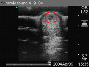
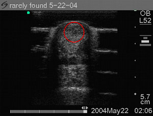
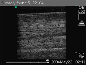
See complete case study details, viewable with Adobe's Acrobat Reader. Authors: Keith Cooper, Elizabeth Bauer, Penny Jenks Initial Clinical Evaluation Rarely Found, a Thoroughbred racehorse colt, was shipped to Penny Jenks, in Louisiana, as a referral from the sate of Florida. Rarely Found was first seen on 4-09-04 by Keith Cooper DVM, for a comprehensive clinical examination that included ultrasonographic imaging. Rarely Found initially presented with a clinical picture of lameness Grade 3.5 out of 5 of the left front limb for one week's duration before diagnosis and treatment. Lameness is graded on a scale of Grade 1 (least lame) to Grade 5 (most lame) in horses. Rarely Found's initial clinical presentation on examination was pain to flexion and palpation of the digital flexor tendon area in about the middle, being, Zone 2. There was well defined, visible swelling as well as heat to the tendon area and edema in the tendon sheath area on palpation. The lesion was positively confirmed by ultrasonographic imaging on the day of the first comprehensive clinical examination and was diagnosed by Keith Cooper DVM as a 25 to 30% core lesion of the superficial digital flexor tendon (SDFT). The lesion was in the Zone 2 A and 2 B area. This core lesion was very concise without evidence of tendon damage out side of the core of the SDFT lesion area. Rehabilitation: Physical Exercise ProgramAt this time, this colt's program of physical activity was confined to stall rest with hand walk for only about 5 minutes for the first 30 days. Initial TreatmentStandard veterinary ancillary treatments included hydrotherapy and icing daily. Standard veterinary inflammation prophylaxis consisted of one injection of hyaluronic acid into the core lesion and tendon sheath. The primary treatment of choice was a protocol of Equine Cymatherapy Bioresonance that consisted of applications of bioresonance to the tendon area as well as to acupuncture points and acupuncture meridians... See complete case study details. Short axis ultrasonography of SDFTdisplaying 25% to 30% core lesion of the superficial digital flexor tendon (SDFT). Six weeks later. Image 2 Ultrasound scan shows a uniform and normal echogenicity with a complete reduction of the SDFT core lesion to a well defined tendon cell regeneration area with no evidence of a prior SDFT lesion. Image 3 shows proper tendon cell and collagen fiber alignment with no further evidence of a prior SDFT core lesion. Continuing Scientific Research This is a short article that examines some of the dynamic bioresonant interactions in the human body in relation to energy fields within the electromagnetic spectrum that we are exposed to on a subatomic and cellular level. Click here to view PDF file. Presentation at 2004 Bioelectromagnetics
Society and Paper on a Recently Anthony H. J. Fleming, Ph.D.,
and Elizabeth Bauer, D.C.B.M., R.N., presented their work of three
years, A predicted photon chemistry, in Washington D.C. at the
annual Bioelectromagnetics Society (BEMS) conference. The BEMS is
the worlds largest scientific society for promoting research and
communication in bioelectromagnetics. Elizabeth Bauer, D.C.B.M., R.N., is director of research, development and education at Cymatherapy International. As a registered nurse, she specialized in critical care medicine before becoming a seeker of greater knowledge and scientific truth in research. As a researcher, she has successfully integrated complementary medical practices into standard medical settings in the United States. She is registered with the World Health Organization as a doctor of cymatics and bioenergetic medicine.
|